Back Of Neck Anatomy Muscles - Pin On Dental School - They move the head in every direction, pulling the skull and jaw towards the shoulders, spine, and scapula.
Back Of Neck Anatomy Muscles - Pin On Dental School - They move the head in every direction, pulling the skull and jaw towards the shoulders, spine, and scapula.. 21 muscles of the neck: The pll starts at c2 and goes down the back of the vertebral bodies and intervertebral discs. William is a final year medical student in australia who has taught anatomy to tertiary science and medical students since 2010. Back muscles are arranged in several layers, so they are divided into deep and superficial, which, in turn, are arranged in two layers. They are divided into three groups, as shown below.
Learn about anatomy neck muscles with free interactive flashcards. The back muscles can be three types. By the middle line of the back is a longitudinal groove back (sulcus dorsi). Spinous processes of txi to liii and supraspinous ligaments. Top head neck anatomy flashcards ranked by quality.
Anatomy Of Back Of Head Anatomy Drawing Diagram from image.slidesharecdn.com The three scalene muscles are found forming the floor of the posterior triangle. Intermediate back muscles and c. There are many muscles around the neck that help to support the cervical spine and allow you to move your head in different directions. The anterior and middle scalenes originate from the transverse processes of certain cervical vertebrae and attach to the first rib. They move the head in every direction, pulling the skull and jaw towards the shoulders, spine, and scapula. Learn about anatomy neck muscles with free interactive flashcards. The muscles of the back that work together to support the spine, help keep the body upright and allow twist and bend in many directions. Neck, face, and intrinsic back muscles.
The back anatomy includes the latissimus dorsi, trapezius, erector spinae, rhomboid, and the teres major.
The major muscle of the back of the neck, the trapezius, is involved in movements of the scapula and is dealt with in the next section, on the muscles in this view of a male figure with one arm up and one arm on the hip, there is a tremendous number of clearly defined anatomical shapes, large and small. There are many muscles around the neck that help to support the cervical spine and allow you to move your head in different directions. Intermediate layer of back muscles. William is a final year medical student in australia who has taught anatomy to tertiary science and medical students since 2010. Adducts, extends and internally rotates the humerus. Short of a great deal of descriptive text, the easiest way to answer this is with illustrations. The neck muscles, including the sternocleidomastoid and the trapezius, are responsible for the gross motor movement in the muscular system of the head and neck. Human muscle system, the muscles of the human body that work the skeletal system, that are under voluntary control, and that the following sections provide a basic framework for the understanding of gross human muscular anatomy, with descriptions of the large muscle groups and their actions. Several other muscles of the back also extend up to the neck region and are partly connected with the cervical part of the vertebral column, including the trapezius, levator scapulae, splenius, iliocostalis, longissimus, rotatores, semispinalis, interspinales, and intertransversarii muscles. Included are views of the back of the neck, short muscles of the neck, prevertebral muscles. The posterior muscles of the neck are primarily concerned with head movements, like extension. The back anatomy includes the latissimus dorsi, trapezius, erector spinae, rhomboid, and the teres major. The muscles of the back that work together to support the spine, help keep the body upright and allow twist and bend in many directions.
Back muscles are arranged in several layers, so they are divided into deep and superficial, which, in turn, are arranged in two layers. In this section, learn more about the anatomy of the muscles of the neck. The back muscles can be three types. This article describes the anatomy of the head and neck of the human body, including the brain, bones, muscles, blood vessels, nerves, glands, nose, mouth, teeth, tongue, and throat. They are divided into three groups, as shown below.
Muscle Anatomy Of The Neck Everything You Need To Know Dr Nabil Ebraheim Youtube from i.ytimg.com They are divided into three groups, as shown below. The back muscles can be three types. Is the only cutaneous muscle in human body (under the skin) attachments: The back muscles stabilize and move the vertebral column, and are grouped according to the lengths and direction of the fascicles. William is a final year medical student in australia who has taught anatomy to tertiary science and medical students since 2010. Working in pairs on the left and. This article describes the anatomy of the head and neck of the human body, including the brain, bones, muscles, blood vessels, nerves, glands, nose, mouth, teeth, tongue, and throat. The splenius muscles originate at the midline and run laterally and.
Neck muscles are bodies of tissue that produce motion in the neck when stimulated. Inserts on to the humerus. They are divided into three groups, as shown below. The muscles of the back that work together to support the spine, help keep the body upright and allow twist and bend in many directions. Border of mandible and skin, and is attached to superficial fascia covering pectoralis major and deltoid muscles inferiorly. Back muscles are arranged in several layers, so they are divided into deep and superficial, which, in turn, are arranged in two layers. Short of a great deal of descriptive text, the easiest way to answer this is with illustrations. Brings down corners of the mouth, expressing. Extrinsic, intermediate and intrinsic muscles. The muscles of the neck run from the base of the skull to the upper back and work together to bend the head and assist in breathing. There are many muscles around the neck that help to support the cervical spine and allow you to move your head in different directions. The major muscle of the back of the neck, the trapezius, is involved in movements of the scapula and is dealt with in the next section, on the muscles in this view of a male figure with one arm up and one arm on the hip, there is a tremendous number of clearly defined anatomical shapes, large and small. The posterior muscles of the neck are primarily concerned with head movements, like extension.
Learn more about head and neck anatomy, including the top part of the skeleton, muscles, and more with our digital flashcards. The muscles of the neck run from the base of the skull to the upper back and work together to bend the head and assist in breathing. Is the only cutaneous muscle in human body (under the skin) attachments: Inserts on to the humerus. Muscles are named according to their shape, location, or a combination.
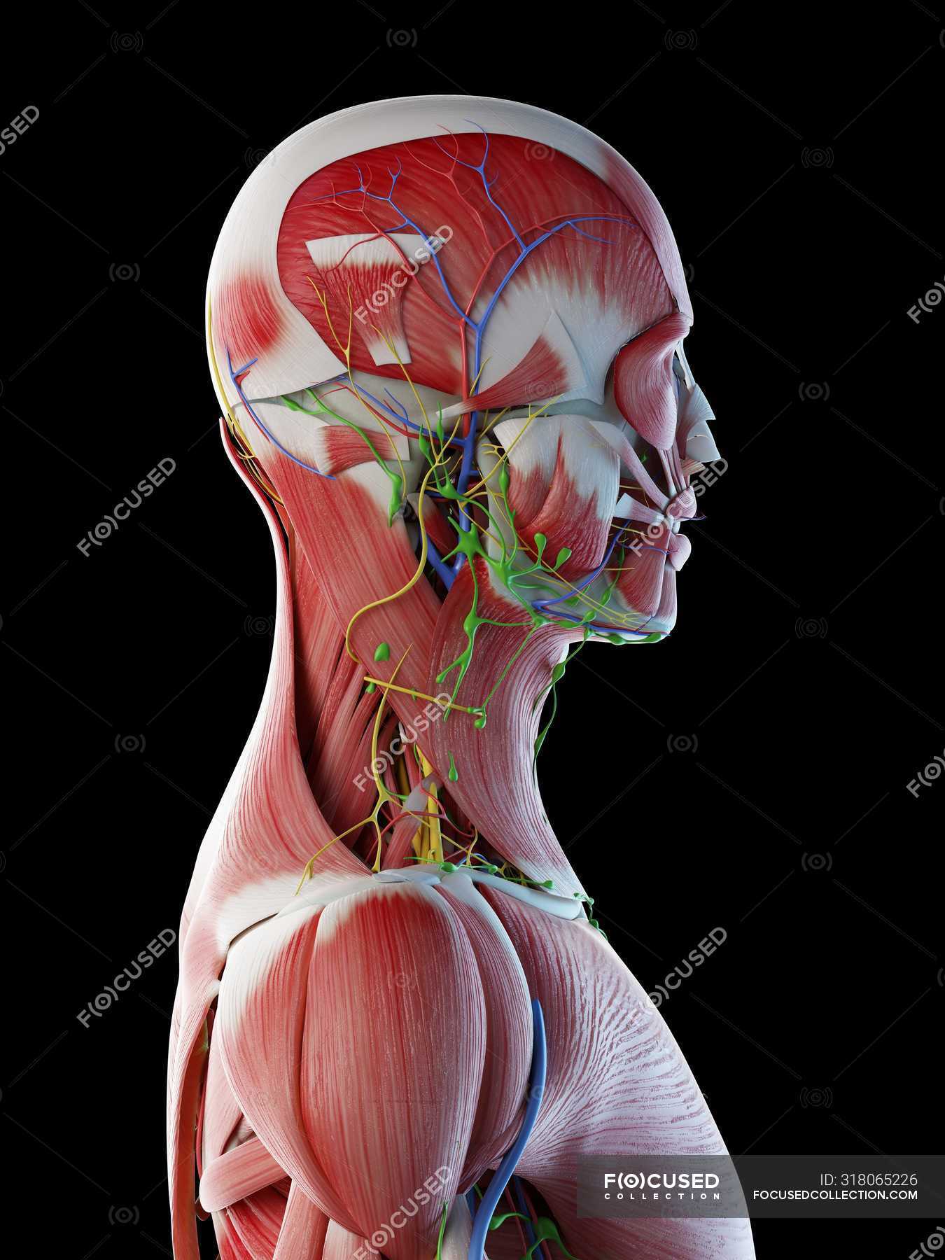
Male Anatomy Of Head Neck And Back With Musculature Computer Illustration Normal Transparent Stock Photo 318065226 from st.focusedcollection.com Intermediate back muscles and c. Neck, face, and intrinsic back muscles. Along it are easily palpable spinous processes by palpation of the cervical vii and all lying. They are further categorized according function such as flexion, extension, or rotation. Bones of the neck picture. Intermediate layer of back muscles. Choose from 500 different sets of flashcards about anatomy neck muscles on quizlet. They are divided into three groups, as shown below.
Intermediate layer of back muscles.
Working in pairs on the left and. Intermediate layer of back muscles. There are several individual muscles within the back anatomy, and it's important to take a quick look the image below to shows all the major back muscles (as well as some neck muscles) Bones of the neck picture. Border of mandible and skin, and is attached to superficial fascia covering pectoralis major and deltoid muscles inferiorly. Whiplash associated disorders and neck rehabilitation. The deep back muscles lie immediately adjacent to the vertebral column and ribs. The back muscles stabilize and move the vertebral column, and are grouped according to the lengths and direction of the fascicles. Anterior muscles of the neck. An online course by chris worsfold. The back has some of the body's largest muscles (erector spinae group) and some of the smallest and most numerous ones. Included are views of the back of the neck, short muscles of the neck, prevertebral muscles. The back muscles can be three types.